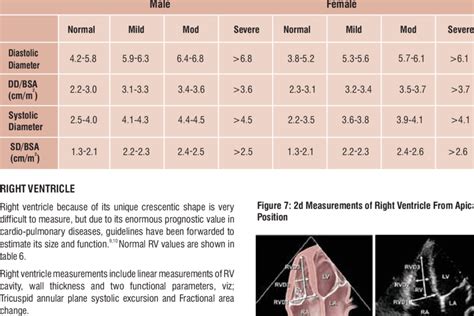
lv height | lv size chart bag lv height This LV mass index calculator helps diagnose the type of cardiac hypertrophy based on LVMI and relative wall thickness. The Donut Bouquet, Las Vegas, Nevada. 3,199 likes. The Original Donut Bouquet Now delivering in Las Vegas & California https://thedonutbouquet.com/.
0 · normal lv size and function
1 · normal lv dimensions
2 · normal lv diameter
3 · lv trainer size chart
4 · lv size normal
5 · lv size chart bag
6 · lv size chart
7 · left ventricle size chart
86K Followers, 2,520 Following, 317 Posts - See Instagram photos and videos from The Dollhouse Las Vegas (@thedollhouselv)
This LV mass index calculator helps diagnose the type of cardiac hypertrophy based on LVMI and relative wall thickness.This LV mass index calculator helps diagnose the type of cardiac hypertrophy based on LVMI and relative wall thickness. Uncontrolled high blood pressure is the most common cause of left ventricular hypertrophy. Complications include irregular heart rhythms, called arrhythmias, and heart failure. Treatment of left ventricular hypertrophy depends on the cause. Treatment may include medications or surgery. To calculate the left ventricular mass index (LVMI), follow these steps: Determine the left ventricular mass using the LV mass equation. Divide the value obtained in step 1 by the body surface area (BSA), calculated based on the patient's weight and .

There are numerous voltage criteria for diagnosing LVH, summarised below. The most commonly used are the Sokolow-Lyon criteria: S wave depth in V 1 + tallest R wave height in V 5 or V 6 > 35 mm. Voltage criteria must be accompanied by non-voltage criteria to be considered diagnostic of LVH. Voltage Criteria. Each echocardiogram includes an evaluation of the LV dimensions, wall thicknesses and function. Good measurements are essential and may have implications for therapy. The LV dimensions must be measured when the end-diastolic and end-systolic valves (MV and AoV) are closed in the parasternal long axis (PLAX) view. Left ventricular hypertrophy (LVH) is a condition in which an increase in left ventricular mass occurs secondary to an increase in wall thickness, an increase in left ventricular cavity enlargement, or both.ECG changes in left ventricular hypertrophy (LVH) and right ventricular hypertrophy (RVH). The electrical vector of the left ventricle is enhanced in LVH, which results in large R-waves in left-sided leads (V5, V6, aVL and I) and deep S-waves in right-sided chest leads (V1, V2).
LVM is the acronym for Left Ventricular Mass. LV mass (LVM) is a vital prognostic measurement we obtain with echocardiography to manage hypertension. RWT is the acronym for Relative Wall Thickness and is an additional reference value that can help further classify the type of LVH.Chapter content. Figure 1. Calculation of left ventricular mass. mass LV = 1.05 (mass total – mass cavity) LV = left ventricle; 1.05 = mycoardial mass constant. Left ventricular hypertrophy (LVH) Left Ventricular Measurement. Left ventricular mass is generally calculated as the difference between the epicardium delimited volume and the left ventricular chamber volume multiplied by an estimate of myocardial density.This LV mass index calculator helps diagnose the type of cardiac hypertrophy based on LVMI and relative wall thickness.
normal lv size and function
Uncontrolled high blood pressure is the most common cause of left ventricular hypertrophy. Complications include irregular heart rhythms, called arrhythmias, and heart failure. Treatment of left ventricular hypertrophy depends on the cause. Treatment may include medications or surgery.
To calculate the left ventricular mass index (LVMI), follow these steps: Determine the left ventricular mass using the LV mass equation. Divide the value obtained in step 1 by the body surface area (BSA), calculated based on the patient's weight and . There are numerous voltage criteria for diagnosing LVH, summarised below. The most commonly used are the Sokolow-Lyon criteria: S wave depth in V 1 + tallest R wave height in V 5 or V 6 > 35 mm. Voltage criteria must be accompanied by non-voltage criteria to be considered diagnostic of LVH. Voltage Criteria.
Each echocardiogram includes an evaluation of the LV dimensions, wall thicknesses and function. Good measurements are essential and may have implications for therapy. The LV dimensions must be measured when the end-diastolic and end-systolic valves (MV and AoV) are closed in the parasternal long axis (PLAX) view. Left ventricular hypertrophy (LVH) is a condition in which an increase in left ventricular mass occurs secondary to an increase in wall thickness, an increase in left ventricular cavity enlargement, or both.ECG changes in left ventricular hypertrophy (LVH) and right ventricular hypertrophy (RVH). The electrical vector of the left ventricle is enhanced in LVH, which results in large R-waves in left-sided leads (V5, V6, aVL and I) and deep S-waves in right-sided chest leads (V1, V2).LVM is the acronym for Left Ventricular Mass. LV mass (LVM) is a vital prognostic measurement we obtain with echocardiography to manage hypertension. RWT is the acronym for Relative Wall Thickness and is an additional reference value that can help further classify the type of LVH.
normal lv dimensions
Chapter content. Figure 1. Calculation of left ventricular mass. mass LV = 1.05 (mass total – mass cavity) LV = left ventricle; 1.05 = mycoardial mass constant. Left ventricular hypertrophy (LVH)
men's burberry crew neck sweater
normal lv diameter
lv trainer size chart
lv size normal
lv size chart bag

georgethecow4 7 years ago #1. http://dragonquest7.nintendo.com/expand/#online-distributions. Nintendo has a list of the current tablets, their distribution dates, and their recommended levels at.
lv height|lv size chart bag


